
Laboratory
The laboratories are geared towards the analysis of carbonate sediments as well as paleoclimates from proxy records using a variety of calcified skeletons of marine biota. However, the setup is versatile enough to allow the examination of a wide range of additional climate archives (e.g., speleothems, tree rings). Our laboratory consists of microanalytical tools, including sample preparation equipment, a fully automated optical imaging system, and microsampling devices. Furthermore, a marine aquaculture laboratory has been outfitted for controlled calibration and growth studies of marine organisms.
Polishing Facility


This facility provides microprobe-quality finish to specimen surfaces up to a size of 15cm. Shown are polished sections of coralline red algae from the North Pacific.
Geo.TS Microscope Mapping System


An automated reflected light microscope (Olympus BX 51) attached to a recently developed sampling stage/imaging system allows 2-dimensional mapping of the surfaces of polished specimens. A motorized large surface XYZ stage (130x85-mm range) and reflected and transmitted light is combined with geo.TS software, which is designed for seamlessly stitching images. A special interface allows overview and zoom functions within an image (similar to the interface used with Google Earth Software). Furthermore, the system can be web-interfaced, so images can be viewed and analyzed from various locations. Click to see coralline red algal virtual image above. For example, growth increment widths of a variety of organisms can be measured using the Olympus 'geo.TS' image analysis software, and used to create annual-resolution climate records ( e.g. more growth = warmer temperatures). The functionality of the recently developed geo.TS system can be viewed at http://www.microscopy.olympus.eu/microscopes/Life_Science_Microscopes__slide.htm (the .slide system shown on this website is comparable to geo.TS)
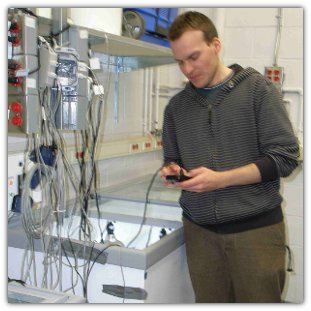
MicroMill


Using the MicroMill (A) we are able to extract minute amounts of carbonate samples (B) along precisely defined drilling paths (C). Samples are later analyzed for stable oxygen isotope composition. (C) shows a section of a coralline red algae from which 500 samples have been extracted.
Aquaculture Laboratory

In our marine aquaculture laboratory we will be monitoring coralline red algae, to be collected in the Northwest Atlantic, as they grow under different light and temperature conditions. A variety of parameters such as temperature, photosynthetically active radiation, salinity, oxygen, pH and calcium are continuously logged or monitored. The outcome of this long-term study is expected to aid linking coralline red algal growth with environmental parameters.
Sample Analysis
Samples prepared in our lab are analyzed in close collaboration with the following analytical facilities.
Oxygen and carbon isotope analysis
Stable Isotope Laboratory (U. Wortmann, University of Toronto) - . Our lab has contributed an autosampler to the Wortmann laboratory for rapid analysis of oxygen and carbon isotopes using a Gas Bench coupled with a Finnigan MAT253 mass spectrometer.
Elemental Analysis
Electron Microprobe Laboratory (Universität Göttingen, Germany, collaboration with A. Kronz) -
Laser Ablation ICP-MS Laboratory (University of Toronto) -
Dating (U/Th)
Multi collector ICP-MS facility (GEOMAR, Germany, collaboration with J. Fietzke) -