Urfa Arain' s Paper on Drosophila Brains & Optic Lobe Visualization
Urfa Arain (PhD student, Erclik lab) and her colleagues (Priscilla Valentino, Ishrat Maliha Islam)’ paper Dissection, Immunohistochemistry and Mounting of Larval and Adult Drosophila Brains for Optic Lobe Visualization was published in Journal of Visualized Experiments
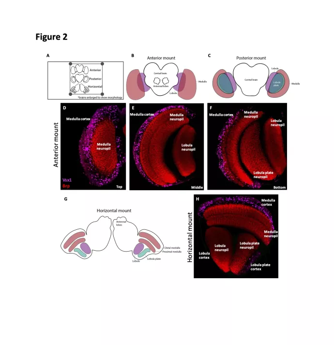
The Drosophila optic lobe, comprised of four neuropils: the lamina, medulla, lobula and lobula plate, is an excellent model system for exploring the developmental mechanisms that generate neural diversity and drive circuit assembly. Given its complex three-dimensional organization, analysis of the optic lobe requires that one understand how its adult neuropils and larval progenitors are positioned relative to each other and the central brain. Here, we describe a protocol for the dissection, immunostaining and mounting of larval and adult brains for optic lobe imaging. Special emphasis is placed on the relationship between mounting orientation and the spatial organization of the optic lobe. We describe three mounting strategies in the larva (anterior, posterior and lateral) and three in the adult (anterior, posterior and horizontal), each of which provide an ideal imaging angle for a distinct optic lobe structure.

Journal of Visualized Experiments : Jove, 28 Apr 2021, (170)
DOI: 10.3791/61273 PMID: 33999033
